Inquiry question: How important is it for genetic material to be replicated exactly?
- Students model the processes involved in cell replication, including but not limited to mitosis and meiosis
Table of Contents
Introduction
One property of living things is their ability to reproduce. In the previous inquiry question, we explored the different strategies used by various organisms to give rise to the next generation. For example, single-celled organisms divide to produce single-celled progeny. Similarly, the progeny of multicellular organisms is also multicellular. However, even multicellular organisms commence life as single cells.
During reproduction, the genetic material is passed on to the progeny. This means that the genetic material in the parental organisms must be copied before it is transferred to the progeny cells. But how faithful is the replicated DNA to the original? The answer depends on the type of reproduction that occurs:
- Asexual reproduction: For cellular divisions such as binary fission and mitosis, the original and copied DNA is nearly identical
- Sexual reproduction: For meiosis, the replicated DNA is a slightly altered version of the original.
The Cell Cycle
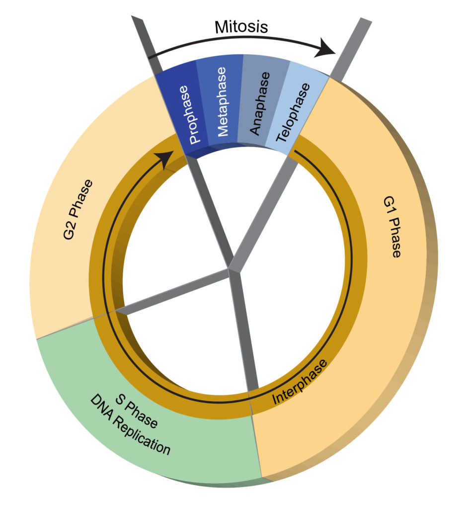
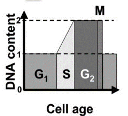
Knowing what happens to cells when not dividing is essential to understanding cell division. The lifetime of a single cell is referred to as its cell cycle. The cell cycle is “a series of events that occurs in a cell as it grows and divides” (NHGRI). The cell cycle is divided into three non-dividing phases, collectively called interphase, and one dividing phase (mitosis or meiosis). The three non-dividing phases of interphase are Synthesis (S) and two rest phases called Gap (G1 and G2). The cell division phase is abbreviated M. This is shown in the following figure.
Mitosis
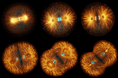
Cells enter mitosis after completing G2. Mitosis is the beginning of cell division. There are two distinct parts to cell division: nuclear division and cytoplasmic division. Nuclear division (mitosis) occurs before cytoplasmic division (cytokinesis). Mitosis consists of four distinct phases based on the behaviours of the chromosomes: prophase, metaphase, anaphase and telophase.
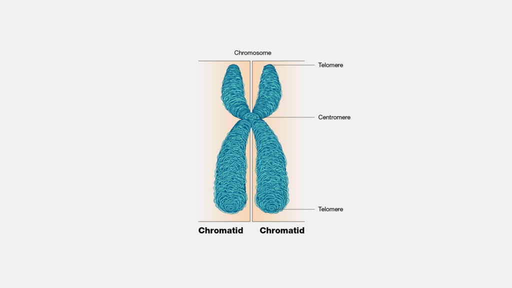
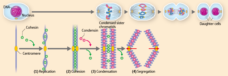
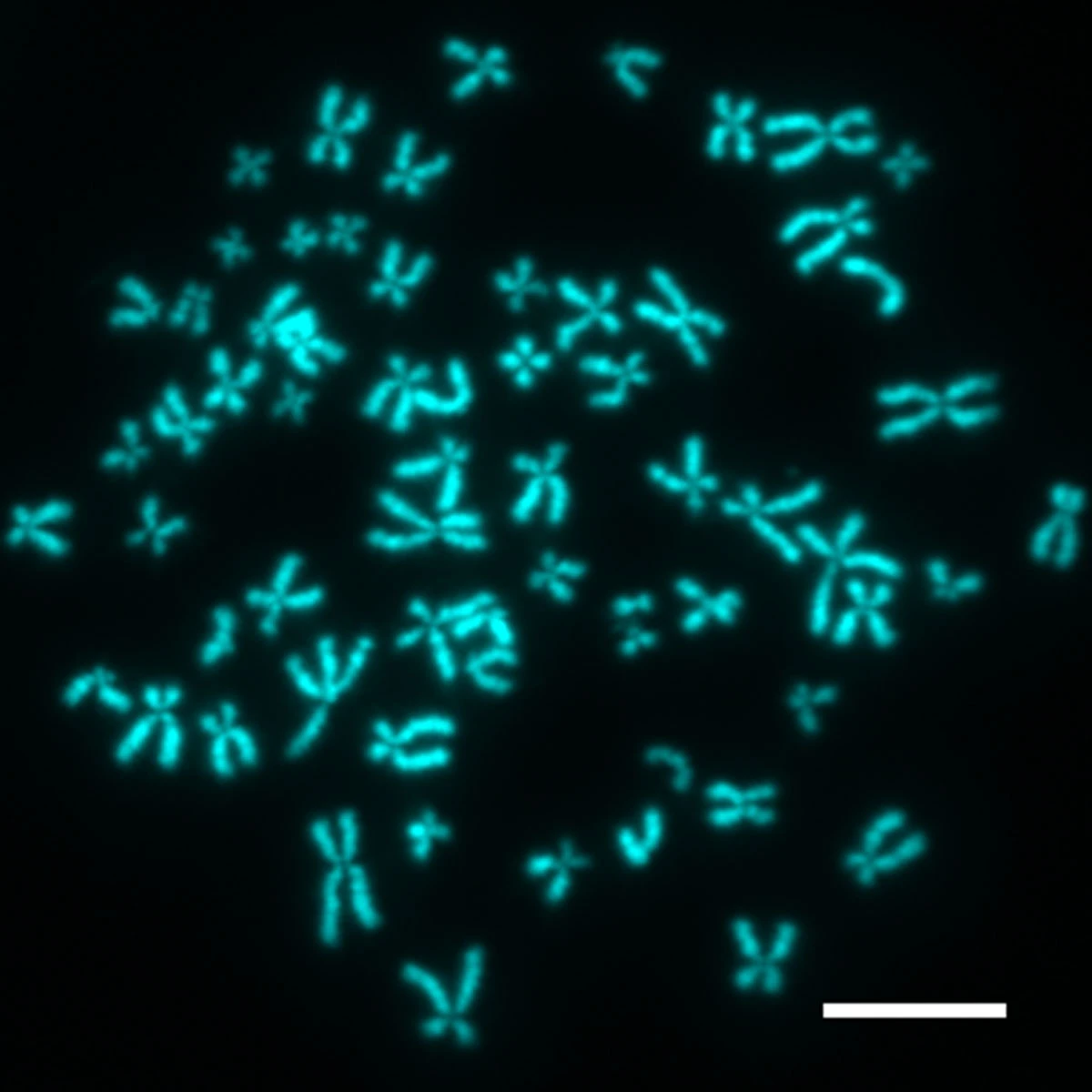
Before the cell enters mitosis, it duplicates its entire genome. For example, a human cell’s genome consists of 46 chromosomes, arranged as 23 pairs. However, when the cell enters mitosis, it has 92 chromosomes (46 x 2). However, they are not arranged as two separate chromosomes but as 46 bivalent chromosomes. The bivalent chromosomes are not visible until the cell is in late prophase.
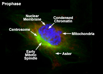
Prophase
Prophase is the first stage of nuclear replication. The main cellular events of prophase are;
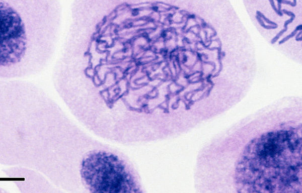
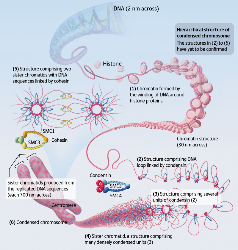
- Condensation of chromosomes: chromosomes become tightly packaged around histone proteins. Condensed chromosomes become visible under the microscope. In addition, condensed chromosomes take up some nuclear stains and appear like short pieces of thread (the word ‘mitosis’ means thread in Greek).
- Duplication of centrioles: centrioles are protein structures that form long protein strands called spindle fibres. The centrioles move to opposite sides of the cell and define the positions of the replicated nuclei. Also, during cytokinesis, the cytoplasm divides in the region between the centrioles.
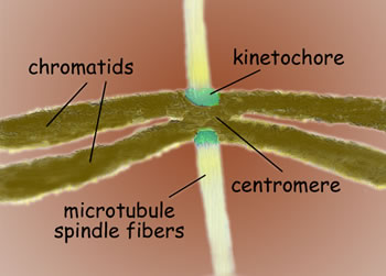
- Spindle fibres: the spindle fibres elongate from the centrioles. They will eventually attach to the chromosomes. This process is so precise that one fibre from each centriole will attach to one chromatid of a bivalent chromosome.
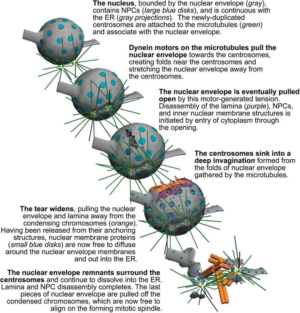
- Breakdown of nuclear envelope: The nuclear envelope is made up of two membranes – that is, it is double-membraned. The membrane disintegrates and forms tiny circular membrane fragments. Those fragments attach to the chromosomes.
By the end of prophase, condensed bivalent chromosomes are seen to be attached to spindle fibres. The growing spindle fibres push the chromosome towards the centre of the cell. This signifies the end of prophase and the start of metaphase.
Nature’s Smart, Elegant Solution to Preserving Genetic Integrity. Science@Berkeley Lab.
Prometaphase
Some authors define the late prophase and early metaphase as a distinct mitotic phase – prometaphase. This article does not discuss prometaphase.
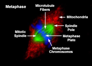
Metaphase
In metaphase, the chromosomes are aligned in the middle of the cell. This region is referred to as the metaphase plate and is approximately halfway between the two centrioles. As mentioned in the previous section, the movement of the chromosomes is caused by the lengthening of the spindle fibres.
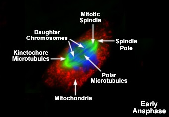
Anaphase
The main event in anaphase is the separation of sister chromatids. The sister chromatids of bivalent chromosomes are held together by proteins that function like glue to hold the replicated DNAs together. At the start of anaphase, specific proteases are activated, resulting in the cleavage of proteins that hold the chromatids together. Then, the spindle fibres contract, pulling the chromatids towards the centrioles. Thus, sister chromatids are separated and move to opposite ends of the cell.
Telophase
Several things occur during telophase.
- Reformation of the nuclear envelope: the membrane fragments associated with the chromosomes fuse. This results informing being formed around the chromosome at the two poles of the cell.
- Decondensation of the chromosomes: The chromosomes will unwrap from the histone proteins (not completely). Individual chromosomes are no longer distinctly visible.
- Dismantling of centrioles and spindle fibres: These structures, which were responsible for separating sister chromatids, are no longer needed by the cell and are dismantled.
By the end of telophase, a cell will have two distinct nuclei.
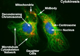
Cytokinesis
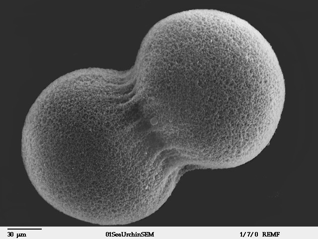
Cytokinesis is not a mitotic phase but a distinct event that occurs after mitosis is completed. Cytokinesis refers to the separation or division of the cytoplasm. This occurs in the region between the two nuclei. Contraction of the cytoskeleton in the middle of the cell (near the metaphase plate) causes the plasma membrane to be pulled to the interior of the cell. This creates a ‘cleavage furrow’. In plants, the cytoplasmic division also includes the laying n of a cellulose cell wall.

Key points
- Genomic duplication occurs before mitosis. In the S-phase of the cell cycle, each DNA molecule is replicated. However, the replicated DNA molecules do not separate but remain attached as sister chromatids.
- After mitosis, each daughter cell has the same number of chromosomes as its parental cell.
- In some organisms, such as some fungal species, cytokinesis does not occur after mitosis. Instead, the two nuclei in the cell will undergo further rounds of mitosis, resulting in large, multi-nucleated cells.
Changes in DNA content during the cell cycle
The amount of DNA a cell changes during the cell cycle. The cell’s ‘normal’ content of DNA is evident during G1. In the S-phase, the DNA content increases because of DNA replication. By the end of the S phase, the cell’s DNA content is double the amount found in the G1 phase. This situation continues until the cell completes G2 and enters mitosis. At the completion of mitosis and cytokinesis, the cell enters G1, and its DNA content returns to ‘normal’.
The role of mitosis
In unicellular eukaryotic organisms, mitosis increases population size. Thus, in those instances, mitosis functions as organismic (whole-organism) reproduction. In multicellular eukaryotes, mitosis contributes to:
- increase in organism size – this is due to the increase in cell numbers in multicellular organisms
- tissue repair and regeneration – mitosis replaces cells that die (apoptosis & necrosis) or are damaged. This often occurs as a result of mitotic divisions of stem cells.